Featured Paper
Integrating brainstem and cortical functional architectures
By Justine Hansen
date: Jan 16 2025
The brainstem needs no introduction. As a fundamental component of the nervous system necessary for survival and consciousness, it is no wonder that any Introduction to Neuroscience textbook will devote many pages on this deep brain structure. However, much of what we know about the brainstem is derived from animal studies and/or ex-vivo histology. As a result, little is known about brainstem function in living humans.
In this study (“Integrating brainstem and cortical functional architectures”), we sought out to understand the functional interplay between the cortex and brainstem in awake humans. This work was possible thanks to recent efforts by the Brainstem Imaging Laboratory at Harvard University in which they not only mapped out 58 brainstem nuclei in MNI152 template space (see the Brainstem Navigator), but also acquired whole-brain resting-state fMRI data using a brainstem-optimized acquisition and preprocessing protocol [1,2]. Here we apply this unique dataset to map out the connection patterns between the whole-brainstem and whole-cortex. We find that:
- Brainstem projections closely map onto cellular architecture (from the BigBrain) and patterns of oscillatory dynamics (from MEG).
- Brainstem nuclei are hierarchically organized into subgroups of nuclei that make distinct cortical projection patterns related to memory, cognitive control, perception, motor coordination, and emotion.
- Using a PET atlas of 18 neurotransmitter receptor/transporter distributions, we find that brainstem-cortex projection patterns are organized according to the neuromodulatory systems in which they participate, and that brainstem nuclei co-localize with receptor and transporter density.
- The putative cortical functional hierarchy – traditionally thought of as a hierarchy of cortico-cortical connectivity – is already present in brainstem-cortex functional connectivity patterns, suggesting that cortical functional organization emerges from brainstem input.
- To ensure all findings are robust, we conduct three sensitivity and robustness analysis where we replicate the analyses (1) using 3 Tesla data acquired in the same individuals, (2) using an alternative parcellation resolution, and (3) in 23 subcortical structures.
Ultimately, we find that multiple cortical phenomena can be traced back to the brainstem, demonstrating the consistent influence that the brainstem exerts on cortical function. This work is published open access in Nature Neuroscience, and is accompanied by a GitHub repository with open code and data.

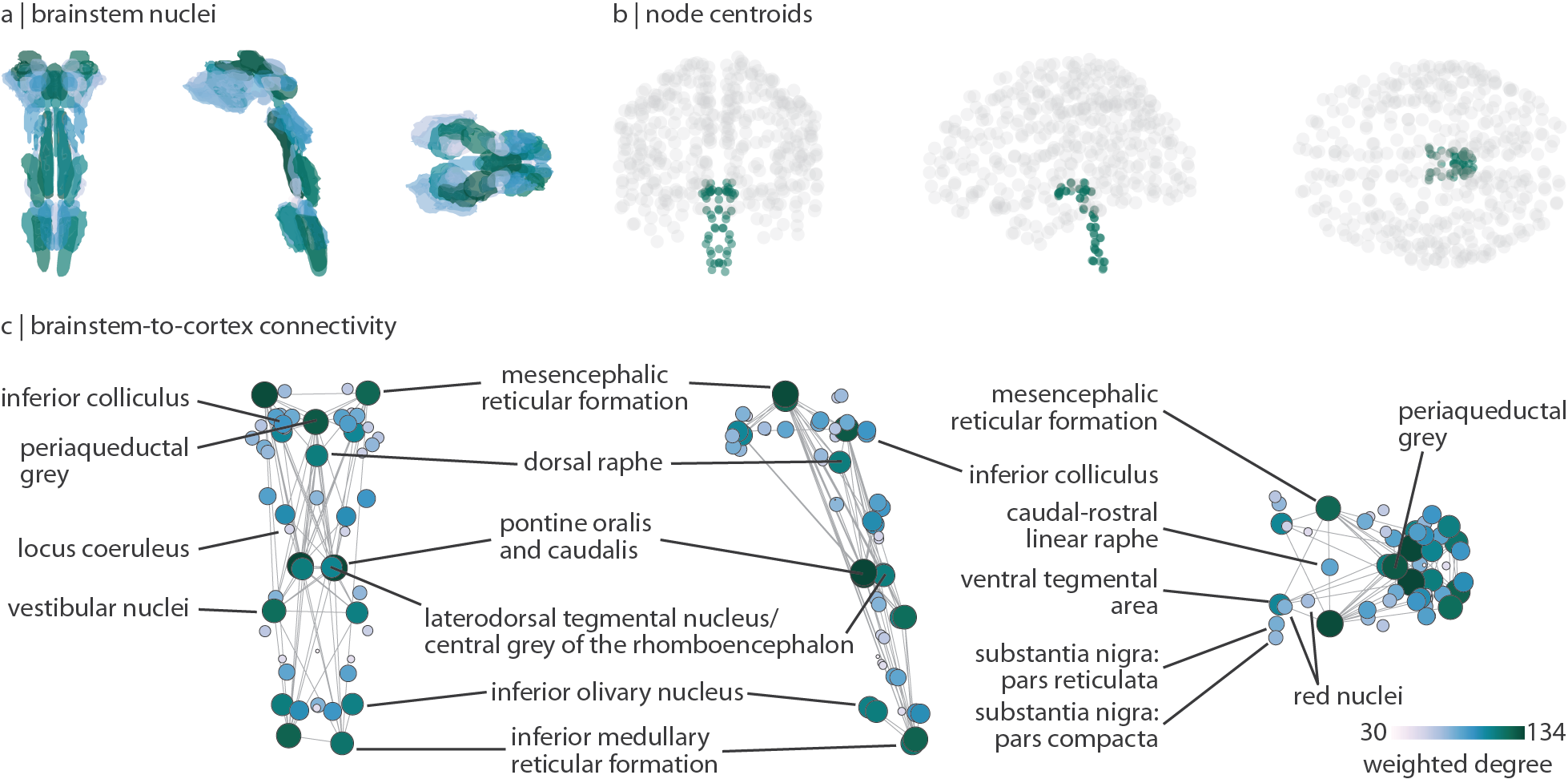
Figure: Extending the connectome to the brainstem. We extend the cortical functional connectome to 58 brainstem nuclei across the midbrain, pons, and medulla using the Brainstem Navigator. (a) Brainstem nuclei (coronal, sagitta, and axial views). (b) Node centroids for the 400 cortical regions and 58 brainstem nuclei used in the main analyses. (c) Weighted degree of brainstem-cortex connectivity (defined as the sum of functional connectivity across all cortical regions for every brainstem nucleus). Key nuclei are labelled.
For more details, see Hansen et al. 2024 Nature Neuroscience (https://www.nature.com/articles/s41593-024-01787-0).
References
- Cauzzo, S., Singh, K., Stauder, M., García-Gomar, M. G., Vanello, N., Passino, C., ... & Bianciardi, M. (2022). Functional connectome of brainstem nuclei involved in autonomic, limbic, pain and sensory processing in living humans from 7 Tesla resting state fMRI. Neuroimage, 250, 118925.
- Singh, K., Cauzzo, S., García-Gomar, M. G., Stauder, M., Vanello, N., Passino, C., & Bianciardi, M. (2022). Functional connectome of arousal and motor brainstem nuclei in living humans by 7 Tesla resting-state fMRI. NeuroImage, 249, 118865.